Imaging in transcatheter native mitral valve replacement with Tendyne mitral valve system: echocardiographic pathway for the interventional imager

All claims expressed in this article are solely those of the authors and do not necessarily represent those of their affiliated organizations, or those of the publisher, the editors and the reviewers. Any product that may be evaluated in this article or claim that may be made by its manufacturer is not guaranteed or endorsed by the publisher.
Authors
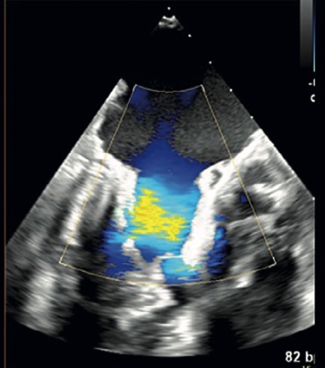
The interaction between the implanter team and the imager team is critical to the success of transcatheter native mitral valve replacement (TMVR), a novel interventional procedure in the therapeutic arsenal for mitral regurgitation. This imaging scenario necessitates the addition of a new dedicated professional figure, dubbed "the interventional imager," with specific expertise in structural heart disease procedures. As its clinical application grows, knowledge of the various imaging modalities used in the TMVR procedure is required for the interventional imager and beneficial for the interventional implanter team. The purpose of this review is to describe the key steps of the procedural imaging pathway in TMVR using the Tendyne mitral valve system, with an emphasis on echocardiography. Pre-procedure cardiac multi-modality imaging screening and planning for TMVR can determine patient eligibility based on anatomic features and measurements, provide measurements for appropriate valve sizing, plan/simulate the access site, catheter/sheath trajectory, and pros- thesis positioning/orientation for correct deployment and predict the risks of potential procedural complications and their likelihood of success. Step-by-step echocardiographic TMVR intraoperative guidance includes: apical access assessment; support for catheter/sheath localization, trajectory and positioning, valve positioning and clocking; post deployment: correct clocking; hemodynamic assessment; detection of perivalvular leakage; obstruction of the left ventricular outlet tract; complications. Knowledge of the multimodality imaging pathway is essential for interventional imagers and critical to the procedure's success.
How to Cite

This work is licensed under a Creative Commons Attribution-NonCommercial 4.0 International License.


