
An unusual case of severe left ventricle outflow tract obstruction due to a coexistence of Takotsubo cardiomyopathy with septal hypertrophic cardiomyopathy
All claims expressed in this article are solely those of the authors and do not necessarily represent those of their affiliated organizations, or those of the publisher, the editors and the reviewers. Any product that may be evaluated in this article or claim that may be made by its manufacturer is not guaranteed or endorsed by the publisher.
Published: 30 December 2021
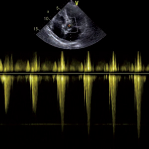
Hypertrophic cardiomyopathy (HCM) is a genetic disorder presenting with a pathological increase of left ventricle (LV) wall thicknesses. The most frequent morphological form is characterized by an abnormal LV basal septal hypertrophy. Tako-Tsubo cardiomyopathy (TTC) is a transient left ventricular systolic dysfunction induced by high physical or emotional stress. Its occurrence with HCM is unusual. However, this presentation occurs more often with the classic asymmetrical septal hypertrophy compared with the apical variant. This case demonstrates that the coexistence of TTC with septal HCM in an elderly patient may lead to a severe hemodinamic instability picture.
Downloads
Kelshiker MA, Mayet J, Unsworth B, Okonko DO. Basal septal hypertrophy. Curr Cardiol Rev 2013;9:325–30.
Armstrong WF, Ryan T, Feigenbaum H. Feigenbaum's Echocardiography. 7th ed. Philadelphia: Wolters Kluwer Health/Lippincott Williams & Wilkins; 2010.
Lee JW, Kim JY. Stress-induced cardiomyopathy: The role of echocardiography. J Cardiovasc Ultrasound 2011;19:7–12.
Turer AT, Samad Z, Valente AM, et al. Anatomic and clinical correlates of septal morphology in hypertrophic cardiomyopathy. Eur J Echocardiogr 2011;12:131–9.
Aboulhosn J, Child JS. Left ventricular outflow obstruction: subaortic stenosis, bicuspid aortic valve, supravalvar aortic stenosis, and coarctation of the aorta. Circulation 2006;114:2412-22.
Nef HM, Möllmann H, Akashi YJ, Hamm CW. Mechanisms of stress (Takotsubo) cardiomyopathy. Nat Rev Cardiol 2010;7:187-93.
De Backer O, Debonnaire P, Gevaert S. et al. Prevalence, associated factors and management implications of left ventricular outflow tract obstruction in takotsubo cardiomyopathy: a two-year, two-center experience. BMC Cardiovasc Disord 2014;14:147.
Afonso L, Bachour K, Awad K, Sandidge G. Takotsubo cardiomyopathy: Pathogenetic insights and myocardial perfusion kinetics using myocardial contrast echocardiography. Eur J Echocardiogr 2008;9:849-54.
El Mahmoud R, Mansencal N, Pilliére R, et al. Prevalence and characteristics of left ventricular outflow tract obstruction in Tako-Tsubo syndrome. Am Heart J 2008;156:543-8.
How to Cite

This work is licensed under a Creative Commons Attribution-NonCommercial 4.0 International License.
PAGEPress has chosen to apply the Creative Commons Attribution NonCommercial 4.0 International License (CC BY-NC 4.0) to all manuscripts to be published.