Comparative analysis of airway invasive aspergillosis and endobronchial spread of tuberculosis on high resolution computed tomography

Submitted: August 22, 2022
Accepted: October 18, 2022
Published: October 25, 2022
Abstract Views: 828
PDF: 0
PDF: 304
Publisher's note
All claims expressed in this article are solely those of the authors and do not necessarily represent those of their affiliated organizations, or those of the publisher, the editors and the reviewers. Any product that may be evaluated in this article or claim that may be made by its manufacturer is not guaranteed or endorsed by the publisher.

Authors

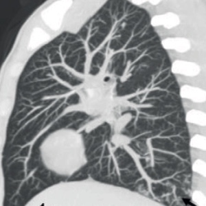
The presence of tree-in-bud (T-I-B) pulmonary opacities on high resolution computed tomography (HRCT) in tuberculosis endemic areas is frequently regarded as a sine qua non for endobronchial tuberculosis (TB). That is not always the case, however. They can also be found in immunocompromised non-neutropenic patients with airway invasive aspergillosis (IA). Understanding the differences between the two conditions is thus critical for making an accurate diagnosis. This research aims to pinpoint those distinguishing characteristics. The study defines the distribution and morphology of T-I-B opacities and other ancillary pulmonary findings in the two conditions by performing a retrospective analysis of HRCT features in 53 immunocompromised patients with lower respiratory tract symptoms, 38 of whom were positive for TB on BAL fluid analysis and 15 confirmed IA by Galactomannan method. While the global distribution of T-I-B opacities affecting all lobes favoured TB (p=0.002), the basal distribution overwhelmingly favoured IA (p<0.0001). Morphologically, dense nodules with discrete margins were associated with TB, whereas nodules with ground-glass density and fuzzy margins were associated with IA. Clustering of nodules was observed in 18 TB patients (p=0.0008). Cavitation was found in 14 (36.84%) of TB patients but not in any of the IA patients. Peri-bronchial consolidation was found in seven (46.67%) of the IA cases and four (10.53%) of the TB cases (p=0.005, 0.007). The presence of ground-glass opacity and bronchiectasis did not differ significantly between the two groups. Not all T-I-B opacities on HRCT chest in immunocompromised patients in endemic TB areas should be reported as tubercular. Immunocompromised non-neutropenic patients with airway IA can be identified earlier with tree-in-bud opacities on HRCT chest, even in the absence of a nodule with halo, resulting in earlier and more effective management.

Dimensions

Altmetric

PlumX Metrics

Downloads

Download data is not yet available.

Citations

Dagenais T, Keller N. Pathogenesis of Aspergillus fumigatus in invasive aspergillosis. Clin Microbiol Rev 2009;22:447-65. DOI: https://doi.org/10.1128/CMR.00055-08
Buckingham S, Hansell D. Aspergillus in the lung: diverse and coincident forms. Eur Radiol 2003;13:1786-800. DOI: https://doi.org/10.1007/s00330-002-1813-4
Lewis R, Kontoyiannis D. Invasive aspergillosis in glucocorticoid-treated patients. Med Mycol 2009;47:S271-81. DOI: https://doi.org/10.1080/13693780802227159
Bassetti M, Peghin M, Vena A. Challenges and Solution of invasive aspergillosis in non-neutropenic patients: A review. Infect Dis Ther 2017;7:17-27. DOI: https://doi.org/10.1007/s40121-017-0183-9
Logan PM, Primack SL, Miller RR, Müller NL. Invasive aspergillosis of the airways: radiographic, CT, and pathologic findings. Radiology 1994;193:383-8. DOI: https://doi.org/10.1148/radiology.193.2.7972747
Greene R, Schlamm H, Oestmann J, et al. Imaging findings in acute invasive pulmonary aspergillosis: Clinical significance of the halo sign. Clin Infect Dis 2007;44:373-9. DOI: https://doi.org/10.1086/509917
Raveendran S, Lu Z. CT findings and differential diagnosis in adults with invasive pulmonary aspergillosis. Radiol Infect Dis 2018;5:14-25. DOI: https://doi.org/10.1016/j.jrid.2018.01.004
Miller W, Panosian J. Causes and imaging patterns of tree-in-bud opacities. Chest. 2013;144:1883-92. DOI: https://doi.org/10.1378/chest.13-1270
Eisenhuber E. The tree-in-bud sign. Radiology 2002;222:771-2. DOI: https://doi.org/10.1148/radiol.2223991980
Vukosav D, Veres KT. Pulmonary tuberculosis in the immunocompromised patients. Vojnosanit Pregl 2019;76:524-30. DOI: https://doi.org/10.2298/VSP170519098V
Youssef J, Novosad S, Winthrop K. Infection risk and safety of corticosteroid use. Rheum Dis Clin North Am 2016;42:157-76. DOI: https://doi.org/10.1016/j.rdc.2015.08.004
Donnelly J, Chen S, Kauffman C, et al. Revision and update of the consensus definitions of invasive fungal disease from the European Organization for Research and Treatment of Cancer and the Mycoses Study Group Education and Research Consortium. Clin Infect Dis 2019;71:1367-76. DOI: https://doi.org/10.1093/cid/ciz1008
Hansell D, Bankier A, MacMahon H, et al. Fleischner Society: Glossary of terms for thoracic imaging. Radiology 2008;246:697-722. DOI: https://doi.org/10.1148/radiol.2462070712
World Health Organization WHO consolidated guidelines on tuberculosis: module 3: diagnosis: rapid diagnostics for tuberculosis detection. Available from: https://www.who.int/publications/i/item/9789240029415
Gupta A, Capoor M, Shende T, et al. Comparative evaluation of galactomannan test with bronchoalveolar lavage and serum for the diagnosis of invasive aspergillosis in patients with hematological malignancies. J Lab Physicians 2017;9:234-8. DOI: https://doi.org/10.4103/JLP.JLP_127_16
Im J, Itoh H. Tree-in-bud pattern of pulmonary tuberculosis on thin-section CT: Pathological implications. Korean J Radiol 2018;19:85. DOI: https://doi.org/10.3348/kjr.2018.19.5.859
Cornillet A, Camus C, Nimubona S et al. Comparison of epidemiological, clinical, and biological features of invasive aspergillosis in neutropenic and nonneutropenic patients: A 6‐year survey. Clin Infect Dis 2006;43:577-84. DOI: https://doi.org/10.1086/505870
Nachiappan A, Rahbar K, Shi X et al. Pulmonary tuberculosis: Role of radiology in diagnosis and management. RadioGraphics 2017;37:52-72. DOI: https://doi.org/10.1148/rg.2017160032

How to Cite

Jamwal, Rupi, Dinesh Singh Kushwaha, Charu Paruthi, Yatish Agarwal, Baljeet Singh Virk, and Malini R. Capoor. 2022. “Comparative Analysis of Airway Invasive Aspergillosis and Endobronchial Spread of Tuberculosis on High Resolution Computed Tomography”. Monaldi Archives for Chest Disease 93 (3). https://doi.org/10.4081/monaldi.2022.2415.