Clinical, radiological and histopathological profile of patients with endobronchial lesions on fibreoptic bronchoscopy

Submitted: May 3, 2022
Accepted: September 13, 2022
Published: September 20, 2022
Abstract Views: 884
PDF: 180
Publisher's note
All claims expressed in this article are solely those of the authors and do not necessarily represent those of their affiliated organizations, or those of the publisher, the editors and the reviewers. Any product that may be evaluated in this article or claim that may be made by its manufacturer is not guaranteed or endorsed by the publisher.

Authors

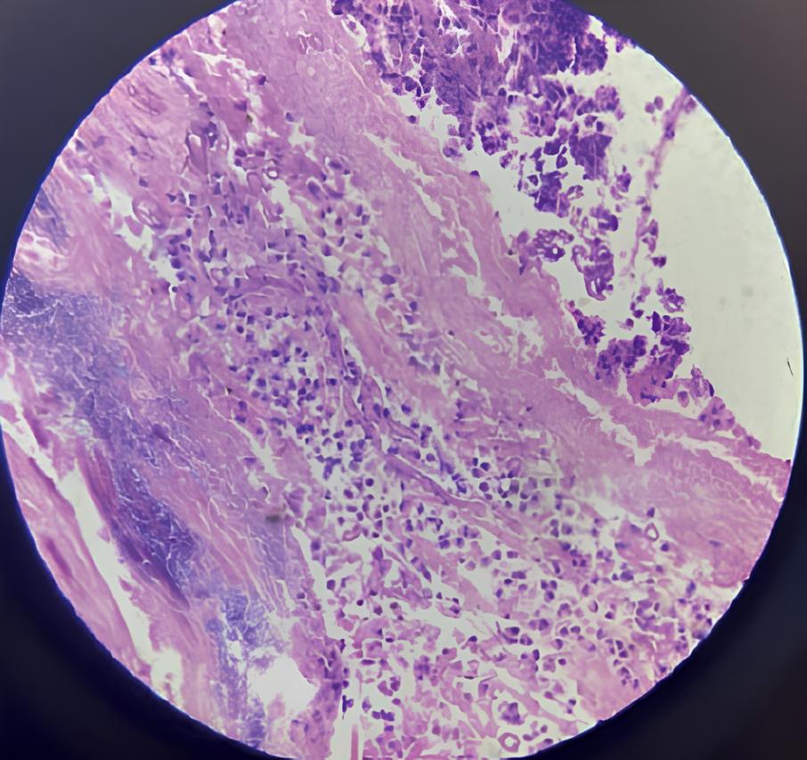
Various pulmonary diseases, both benign as well as malignant, manifest in the form of endobronchial lesions on bronchoscopy. Malignancy is frequently the provisional diagnosis in the mind of a chest physician undergoing an endobronchial biopsy. Other benign diseases, however, may present similarly on bronchoscopy and computerized tomography (CT) scan. This observational study was conducted to better understand why there is such a wide range of endobronchial lesions with even more diverse radiological and pathological presentations. The research was carried out at the Department of Respiratory Medicine, Himalayan Institute of Medical Science (HIMS), Swami Ram Nagar, Dehradun. Subjects were recruited from HIMS, Dehradun patients over a 12-month period (August 2020 to July 2021). The study included patients (over the age of 18) who had a fibreoptic bronchoscopy and were found to have an endobronchial lesion. After a thorough history, examination, and application of the inclusion and exclusion criteria, 120 patients were enrolled. The majority of patients were between the ages of 56 and 65, with males outnumbering females. The majority of the patients were smokers, and the most common complaint was shortness of breath. Poorly differentiated carcinoma and squamous cell carcinoma were the most common endobronchial lesions in men, while small cell carcinoma was the most common in women. A mass lesion was the most common radiological finding, followed by mediastinal lymphadenopathy, and an exophytic lesion was the most common endobronchial lesion detected in bronchoscopy. We looked at the diseases that cause endobronchial lesions and their clinico-radiological and histopathological profiles. This study clearly demonstrates the importance of studying the histopathological profiles of patients with endobronchial growth, which can mimic malignancy in rare cases.

Dimensions

Altmetric

PlumX Metrics

Downloads

Download data is not yet available.

Citations

Wilson RW, Kirejczyk W. Pathological and radiological correlation of endobronchial neoplasms: Part I, Benign tumors. Ann Diagn Pathol 1997;1:31-46. DOI: https://doi.org/10.1016/S1092-9134(97)80007-X
Wilson RW, Frazier AA. Pathological and radiological correlation of endobronchial neoplasms: part II, malignant tumors. Ann Diagn Pathol 1998;2:31-54. DOI: https://doi.org/10.1016/S1092-9134(98)80033-6
Kumar T, Epstein M, Markovskaya Y, et al. Bronchoscopy and endobronchial disease in patients with human immunodeficiency virus infection. Indian J Chest Dis Allied Sci 2011;53:99.
Jin SL, Lee HP, Kim JI, et al. A case of endobronchial actinomycosis. Korean J Intern Med 2000;15:240. DOI: https://doi.org/10.3904/kjim.2000.15.3.240
Kim JS, Rhee Y, Kang SM, et al. A case of endobronchial aspergilloma. Yonsei Med J 2000;41:422-5. DOI: https://doi.org/10.3349/ymj.2000.41.3.422
Donohue JF. Endobronchial mucormycosis. Chest 1983;83:585. DOI: https://doi.org/10.1378/chest.83.3.585b
Hause DW, Harvey JC. Endobronchial carcinoid and mucoepidermoid carcinoma in children. J Surg Oncol 1991;46:270-2. DOI: https://doi.org/10.1002/jso.2930460412
Nilsson JR, Restrepo CS, Jagirdar J. Two cases of endobronchial carcinoid masked by superimposed aspergillosis: a review of the literature of primary lung cancers associated with Aspergillus. Ann Diagn Pathol 2013;17:131-6. DOI: https://doi.org/10.1016/j.anndiagpath.2011.06.005
Corona FE, Okeson GC. Endobronchial fibroma: An unusual case of segmental atelectasis. Am Rev Respir Dis 1974;110:350-3. DOI: https://doi.org/10.1164/arrd.1974.110.3.350
Rose AS, Mathur PN. Endobronchial capillary hemangioma: case report and review of the literature. Respiration 2008;76:221-4. DOI: https://doi.org/10.1159/000099008
Hollings N, Shaw P. Diagnostic imaging of lung cancer. Eur Respir J 2002;19:722-42. DOI: https://doi.org/10.1183/09031936.02.00280002
Toyoda Y, Nakayama T, Kusunoki Y, et al. Sensitivity and specificity of lung cancer screening using chest low-dose computed tomography. Br J Cancer 2008;98:1602-7. DOI: https://doi.org/10.1038/sj.bjc.6604351
Lubell DL. Drawbacks and limitations of computed tomography. Tex Heart Inst J 2005;32:250.
Ikeda N, Hayashi A, Iwasaki K, et al. Comprehensive diagnostic bronchoscopy of central type early-stage lung cancer. Lung Cancer 2007;56:295-302. DOI: https://doi.org/10.1016/j.lungcan.2007.01.009
Rivera MP, Mehta AC, Wahidi MM. Establishing the diagnosis of lung cancer: Diagnosis and management of lung cancer, 3rd ed: American College of Chest Physicians evidence-based clinical practice guidelines. Chest 2013;143:e142S-65S. DOI: https://doi.org/10.1378/chest.12-2353
Rana S, Bhattacharyya BD, Katoch CD, et al. Clinical, radiological, and histopathological profile of patients with endobronchial lesions on fiber-optic bronchoscopy. J Assoc Chest Phys 2018;6:53. DOI: https://doi.org/10.4103/jacp.jacp_27_17
Halima KM, Makled SF, Basiony FS. Histopathological patterns of endobronchial lesions and the role of flexible fiberoptic bronchoscopy in their diagnosis. Al-Azhar Assiut Med J 2020;18:284-9. DOI: https://doi.org/10.4103/AZMJ.AZMJ_39_19
Sharma A, Gandotra N. Role of fiberoptic bronchoscopy in establishing the diagnosis of various lung diseases in a tertiary care hospital. int J Sci Res 2015;4:1498-500.
Devkota KC, Pathak R, Khanal A, Chokhani R. Fiber-optic bronchoscopy: seven-year experience at Nepal Medical College Teaching Hospital. Nepal Med Coll J 2010;12:260-3.
Ma JE, Yun EY, Kim YE, et al. Endobronchial aspergilloma: report of 10 cases and literature review. Yonsei Med J 2011;52:787-92. DOI: https://doi.org/10.3349/ymj.2011.52.5.787
U.S. Department of Health and Human Services. The health consequences of involuntary exposure to tobacco smoke; a report of the Surgeon General. Available from: https://stacks.cdc.gov/view/cdc/13252/
Pavlovska I, Danilovski D, Orovchanec N, et al. An epidemiologic study of some characteristics of lung cancer. Folia Medica 2004;423-31.
Alamoudi OS. Lung cancer at a university hospital in Saudi Arabia: a four-year prospective study of clinical, pathological, radiological, bronchoscopic, and biochemical parameters. Ann Thorac Med 2010;5:30. DOI: https://doi.org/10.4103/1817-1737.58957
Rabahi MF, Ferreira AA, Reciputti BP, et al. Fiberoptic bronchoscopy findings in patients diagnosed with lung cancer. J Bras Pneumol 2012;38:445-51. DOI: https://doi.org/10.1590/S1806-37132012000400006
Devesa SS, Bray F, Vizcaino AP, Parkin DM. International lung cancer trends by histologic type: male: female differences diminishing and adenocarcinoma rates rising. Int J Cancer 2005;117:294-9. DOI: https://doi.org/10.1002/ijc.21183
Husari AW, Jensen WA, Kirsch CM, et al. Pulmonary mucormycosis presenting as an endobronchial lesion. Chest 1994;106:1889-91. DOI: https://doi.org/10.1378/chest.106.6.1889
Hurt RA, Bates MI. Carcinoid tumours of the bronchus: a 33-year experience. Thorax 1984;39:617-23. DOI: https://doi.org/10.1136/thx.39.8.617
Fink G, Krelbaum T, Yellin A, et al. Pulmonary carcinoid: presentation, diagnosis, and outcome in 142 cases in Israel and review of 640 cases from the literature. Chest 2001;119:1647-51. DOI: https://doi.org/10.1378/chest.119.6.1647
Shahzad T, Irfan M. Endobronchial tuberculosis - a review. J Thorac Dis 2016;8:3797. DOI: https://doi.org/10.21037/jtd.2016.12.73
Insler JE, Seder CW, Furlan K, et al. Benign endobronchial tumors: A clinicopathologic review. Front Surg 2021;8:644656. DOI: https://doi.org/10.3389/fsurg.2021.644656

Ethics Approval

The study protocol was approved by the Ethical Review Committee of the Swami Rama Himalayan University

How to Cite

Sharma, Deepen, Rakhee Khanduri, Shailendra Raghuvanshi, Smita Chandra, Sushant Khanduri, Varuna Jethani, and Manoj Kumar. 2022. “Clinical, Radiological and Histopathological Profile of Patients With Endobronchial Lesions on Fibreoptic Bronchoscopy”. Monaldi Archives for Chest Disease 93 (3). https://doi.org/10.4081/monaldi.2022.2312.