
Cardiology - Case Reports
Vol. 93 No. 1 (2023)
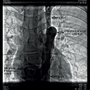
Hemiazygous continuation of inferior vena cava draining into the coronary sinus via persistent left superior vena cava: a rare anomaly
Publisher's note
All claims expressed in this article are solely those of the authors and do not necessarily represent those of their affiliated organizations, or those of the publisher, the editors and the reviewers. Any product that may be evaluated in this article or claim that may be made by its manufacturer is not guaranteed or endorsed by the publisher.
All claims expressed in this article are solely those of the authors and do not necessarily represent those of their affiliated organizations, or those of the publisher, the editors and the reviewers. Any product that may be evaluated in this article or claim that may be made by its manufacturer is not guaranteed or endorsed by the publisher.
Received: 26 March 2022
Accepted: 18 June 2022
Accepted: 18 June 2022
1510
Views
1116
Downloads
Authors
We present a case of left sided inferior vena cava with hemiazygous continuation draining into the coronary sinus via the left persistent superior vena cava. This was incidentally found in an individual referred to our centre for evaluation of palpitations. These caval anomalies are rare, and are often associated with no clinical manifestations. However, it is necessary to recognize them during routine workup to avoid diagnostic and procedural pitfalls.
Downloads
Download data is not yet available.
Citations


Dahnert W. In: 2nd ed, Radiology review manual, Baltimore: Williams and Wilkins; 1996, p. 433.
Bass JE, Redwine MD , Kramer LA, et al . Spectrum of congenital anomalies of the inferior vena cava: cross-sectional imaging findings. Radiographics 2000;20:639-52. DOI: https://doi.org/10.1148/radiographics.20.3.g00ma09639
Sonavane S, Milner D, Singh S, et al. Comprehensive imaging review of the superior vena cava. Radiographics 2015;35:1873-92. DOI: https://doi.org/10.1148/rg.2015150056
Knudtzon J, Svane S. Left sided inferior vena cava. Acta Chir Scand 1986;152:547–9.
Alharthi M, Mookadam F, Collins J, et al. Images in cardiovascular medicine. Extracardiac venous heterotaxy syndrome: complete noninvasive diagnosis by multimodality imaging. Circulation 2008;117:e498-503. DOI: https://doi.org/10.1161/CIRCULATIONAHA.107.741041
Ojha V, Pandey NN, Jagia P. Hemiazygos continuation of isolated left-sided inferior vena cava into persistent left superior vena cava: rare association of left isomerism. BMJ Case Rep 2019;12:e230350. DOI: https://doi.org/10.1136/bcr-2019-230350
Kabakus I, Kocher M, Agha A, Burt JR. Left-sided inferior vena cava with hemiazygos continuation to left superior vena cava. Cureus 2019;11:e6503. DOI: https://doi.org/10.7759/cureus.6503
Kim HJ, Ahn IO, Park ED. Hemiazygos continuation of a left inferior vena cava draining into the right atrium via persistent left superior vena cava: demonstration by helical computed tomography. Cardiovasc Intervent Radiol 1995;18:65-7. DOI: https://doi.org/10.1007/BF02807362
Kim YJ, Kwon HS, Ahn SE et al. Interrupted inferior vena cava with hemiazygos continuation in an adult with a persistent left superior vena cava and left single coronary artery: A case report. J Korean Soc Radiol 2016;74:394-8. DOI: https://doi.org/10.3348/jksr.2016.74.6.394
Freedom RM. Angiography of congenital heart disease, London: Collier Macmillan; 1984, pp. 46-61.
Kraimps JL, Dib H, Raynier P, et al. Left-sided inferior vena cava and thrombosis: case report. Eur J Surg 1993;159:441-3.
Brener BJ, Darling RC, Frederic PL, et al. Major venous anomalies complicating abdominal aortic surgery. Arch Surg 1974;108:159-65. DOI: https://doi.org/10.1001/archsurg.1974.01350260019004
How to Cite
“Hemiazygous Continuation of Inferior Vena Cava Draining into the Coronary Sinus via Persistent Left Superior Vena Cava: A Rare Anomaly”. 2022. Monaldi Archives for Chest Disease 93 (1). https://doi.org/10.4081/monaldi.2022.2275.
Copyright (c) 2022 The Author(s)


This work is licensed under a Creative Commons Attribution-NonCommercial 4.0 International License.