Use of flow volume curve to evaluate large airway obstruction

Submitted: May 25, 2021
Accepted: January 16, 2022
Published: February 4, 2022
Abstract Views: 1727
PDF: 1072
Publisher's note
All claims expressed in this article are solely those of the authors and do not necessarily represent those of their affiliated organizations, or those of the publisher, the editors and the reviewers. Any product that may be evaluated in this article or claim that may be made by its manufacturer is not guaranteed or endorsed by the publisher.

Authors

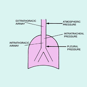
The flow volume loop (FVL) is a graphic display of airflow against lung volumes at different levels obtained during the maximum inspiratory and expiratory maneuver. It is a simple and reproducible method of lung function assessment. A narrative review was performed according to the Preferred Reporting Items for Systematic Reviews and Meta-Analysis (PRISMA) guidelines. PubMed, EMBASE, Ovid MEDLINE and CINAHL databases were queried and reviewed for studies pertinent to the various FVLs abnormalities and their mechanisms from January 2020 to December 2020. We used the following search terms; flow-volume loop, upper airway obstruction, Obstructive airway disease, and spirometry.  Assessing the shape of the flow-volume loop is particularly helpful in diagnosing and localizing upper airway obstruction. They are also helpful in identifying bronchodilator response to treatment. Characteristic FVLs is also seen in patients with obstructive or restrictive lung disorders. Spirometry should be interpreted using the absolute values for flows and volumes as well as the flow volume and volume time curves.

Dimensions

Altmetric

PlumX Metrics

Downloads

Download data is not yet available.

Citations

Hyatt RE, and Black LF. The flow-volume curve. A current perspective. Am Rev Respir Dis 1973;107:191-9.
Pellegrino R, Viegi G, and Brusasco V, et al. Interpretative strategies for lung function tests. Eur Respir J 2005;26:948-68. DOI: https://doi.org/10.1183/09031936.05.00035205
Miller RD and Hyatt RE. Evaluation of obstructing lesions of the trachea and larynx by flow-volume loops. Am Rev Respir Dis 1973;108:475-41.
Moore VC. Spirometry: step by step. Breath 2012;8:233-40. DOI: https://doi.org/10.1183/20734735.0021711
Kryger M, Bode F, Antic R, et al. Diagnosis of obstruction of upper and central airways. Am J Med 1976;61:85-93. DOI: https://doi.org/10.1016/0002-9343(76)90048-6
Lunn WW, Sheller JR. Flow volume loops in the evaluation of upper airway obstruction. Otolaryngol Clin North Am 1995;28:721-9. DOI: https://doi.org/10.1016/S0030-6665(20)30490-4
Martin L. Pulmonary physiology in clinical practice: the essentials for patient care and evaluation. St Louis: Mosby Inc.;1987.
Mead J, Turner JM, Macklem PT, et al. Significance of the relationship between lung recoil and maximum expiratory flow. J Appl Physiol 1967;22:95-108. DOI: https://doi.org/10.1152/jappl.1967.22.1.95
Empey DW: Assessment of upper airway obstruction. Br Med J 1972;3:503-5. DOI: https://doi.org/10.1136/bmj.3.5825.503
Miller RD, Hyatt RE. Obstructing lesions of the larynx and trachea: clinical and physiologic characteristics. Mayo Clin Proc 1969;44:145-61.
Raposo LB, Bugalho A, Gomes MJ. Contribution of flow-volume curves to the detection of central airway obstruction. J Bras Pneumol 2013;39:447-54. DOI: https://doi.org/10.1590/S1806-37132013000400008
Acres JC, Kryger MH. Clinical significance of pulmonary function tests: upper airway obstruction. Chest 1981;80:207-11. DOI: https://doi.org/10.1378/chest.80.2.207
Yernault Je, Englert M, and Sergysels R, De Coster A. Upper airway stenosis: A physiologic study. Am Rev Respir Dis 1973;108:999-1000.
Stoller JK. Spirometry: a key diagnostic test in pulmonary medicine. Cleve Clin J Med 1992;59:75-8. DOI: https://doi.org/10.3949/ccjm.59.1.75
Sterner JB, Morris MJ, Sill JM, Hayes JA. Inspiratory flow-volume curve evaluation for detecting upper airway disease. Respir Care 2009; 54:461-6.
Bolliger CT, Sofko J, Maurer P, Perruchoud AP. The flow-volume loop in bilateral vocal cord paralysis. Chest 1993;104:1302-4. DOI: https://doi.org/10.1378/chest.104.4.1302
Bahrainwala AH, Simon MR, Harrison DD, et al. Atypical expiratory flow volume curve in an asthmatic patient with vocal cord dysfunction. Ann Allergy Asthma Immunol 2001;86:439-43. DOI: https://doi.org/10.1016/S1081-1206(10)62492-1
Guntupalli KK, Bandi V, Sirgi C, et al. Usefulness of flow volume loops in emergency center and ICU settings. Chest 1997;111:481-8. DOI: https://doi.org/10.1378/chest.111.2.481
Hyatt RE. Evaluation of major airway lesions using the flow-volume loop. Ann Otol Rhinol Laryngol 1975;84:635-42. DOI: https://doi.org/10.1177/000348947508400514
Aboussouan LS, Stoller JK. Diagnosis and management of upper airway obstruction. Clin Chest Med 1994;15:35-53. DOI: https://doi.org/10.1016/S0272-5231(21)01054-6
Gelb AF, Tashkin DP, Epstein JD, et al. Physiologic characteristics of malignant unilateral mainstem bronchial obstruction: diagnosis and Nd-YAG laser treatment. Am Rev Respir Dis 1988;138:1382-5. DOI: https://doi.org/10.1164/ajrccm/138.6.1382
Gascoigne AD, Corris PA, Dark JH, Gibson GJ. The biphasic spirogram: a clue to unilateral narrowing of a mainstem bronchus. Thorax 1990;45:637-8. DOI: https://doi.org/10.1136/thx.45.8.637
Anzueto A, Levine SM, Tillis WP, et al. Use of flow volume loop in the diagnosis of bronchial stenosis after single lung transplantation. Chest 1994;105:934–6. DOI: https://doi.org/10.1378/chest.105.3.934
Neagos GR, Martinez FJ, and Deeb GM, et al. Diagnosis of unilateral mainstem bronchial obstruction following single-lung transplantation with routine spirometry. Chest 1993;103:1255-8. DOI: https://doi.org/10.1378/chest.103.4.1255
Roos CM, Braat MC. End-inspiratory flow reduction in the forced flow-volume curve as a sign of unilateral bronchial disease. Neth J Med 1985;28:378-82.
Agrawal A, Sahni S, Marder G, et al. Biphasic flow-volume loop in granulomatosis with polyangiitis related unilateral bronchus obstruction. Respir Investig 2016;54:280-3. DOI: https://doi.org/10.1016/j.resinv.2016.01.002
Jeong YG, Donohue JF. Unilateral bronchial occlusion in a patient with sarcoidosis and a bronchial anomaly. South Med J 1980;73:523–4. DOI: https://doi.org/10.1097/00007611-198004000-00036
Majid A, Sosa AF, Ernst A, et al. Pulmonary function and flow- volume loop patterns in patients with tracheobronchomalacia. Respir Care 2013;58:1521–6. DOI: https://doi.org/10.4187/respcare.02277
Sanders MH, Martin RJ, Pennock BE, Rogers RM. The detection of sleep apnea in the awake patient: The 'saw tooth' sign. JAMA 1981;245:2414-18. DOI: https://doi.org/10.1001/jama.245.23.2414
Schiffman PL. A "saw-tooth" pattern in Parkinson's disease. Chest 1985;87:124-6. DOI: https://doi.org/10.1378/chest.87.1.124
Vincken WG, Cosio MG. Flow oscillations on the flow-volume loop: clinical and physiological implications. Eur Respir J 1989;2:543-9.
Vincken WG, Gauthier SG, Dollfuss RE, et al. Involvement of upper-airway muscles in extrapyramidal disorders. A cause of airflow limitation. N Engl J Med 1984;311:438-42. DOI: https://doi.org/10.1056/NEJM198408163110704
Garcia-Pachon E. Tracheobronchomalacia: a cause of flow oscillations on the flow-volume loop. Chest 2000;118:1519. DOI: https://doi.org/10.1378/chest.118.5.1519
Das N, Topalovic M, Aerts JM, et al. Area under the forced expiratory flow-volume loop in spirometry indicates severe hyperinflation in COPD patients. Int J Chron Obstruct Pulmon Dis 2019;14:409-18. DOI: https://doi.org/10.2147/COPD.S185931
Langer D, Ciavaglia CE, Neder JA, et al. Lung hyperinflation in chronic obstructive pulmonary disease: mechanisms, clinical implications and treatment. Expert Rev Respir Med 2014;8:731-49. DOI: https://doi.org/10.1586/17476348.2014.949676
Campbell AH, Faulks LW. Expiratory airflow patterns in tracheobronchial collapse. Am Rev Respir Dis 1965;92:781-91.
Gandevia B. The spirogram of gross expiratory tracheobronchial collapse in emphysema. Q J Med 1963;32:23-31.
Jayamanne DS, Epstein H, Goldring RM. Flow volume curve contour in COPD: correlation with pulmonary mechanics. Chest 1980;77:749–57. DOI: https://doi.org/10.1378/chest.77.6.749
Murgu SD, Colt HG. Tracheobronchomalacia and excessive dynamic airway collapse. Respirology 2006;11:388-406. DOI: https://doi.org/10.1111/j.1440-1843.2006.00862.x
Karkhanis VS, Joshi JM. Spirometry in chronic obstructive lung disease (COPD). J Assoc Physicians India 2012;60 Suppl:22-6.
Arora VK, Raghu S. Flow volume curves: clinical significance. Lung India 1996;XIV: 169-71.
Vincken W, Elleker G, Cosio MG. Detection of upper airway muscle involvement in neuromuscular disorders using the flow-volume loop. Chest 1986;90:52-7. DOI: https://doi.org/10.1378/chest.90.1.52
Vincken WG, Elleker MG, Cosio MG. Flow–volume loop changes reflecting respiratory muscle weakness in chronic neuromuscular disorders. Am J Med 1987;83:673-80. DOI: https://doi.org/10.1016/0002-9343(87)90897-7
Hancox B, Whyte K. McGraw-Hill’s pocket guide to lung function tests. North Ryde: McGraw-Hill; 2006.
Hyatt RE, Scanlon PD, Nakamura M. Interpretation of pulmonary function tests: A practical guide. Philadelphia: Lippincott-Raven; 2007.
Shin HH, Sears MR, Hancox RJ. Prevalence and correlates of a 'knee' pattern on the maximal expiratory flow-volume loop in young adults. Respirology 2014;19:1052. DOI: https://doi.org/10.1111/resp.12352
Mead J. Expiratory flow limitation: a physiologist’s point of view. Fed Proc 1980;39:2771-5.

How to Cite

Sarkar, Malay, Irappa V. Madabhavi, Shailee Mehta, and Swadeshkumar Mohanty. 2022. “Use of Flow Volume Curve to Evaluate Large Airway Obstruction”. Monaldi Archives for Chest Disease 92 (4). https://doi.org/10.4081/monaldi.2022.1947.