Pulmonary actinomycosis: cytomorphological features

Submitted: October 15, 2020
Accepted: September 22, 2021
Published: November 5, 2021
Abstract Views: 1117
PDF: 670
Publisher's note
All claims expressed in this article are solely those of the authors and do not necessarily represent those of their affiliated organizations, or those of the publisher, the editors and the reviewers. Any product that may be evaluated in this article or claim that may be made by its manufacturer is not guaranteed or endorsed by the publisher.

Authors

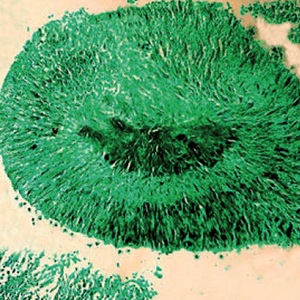
Pulmonary actinomycosis is an uncommon infectious disease. Although the gold standard for diagnosis is histological examination with bacterial culture of lung tissue, cytology samples offer a fast and low-cost alternate diagnostic procedure. The cytology literature on this topic is limited to mostly case reports. Therefore, the aim of this study was to review cytological material in a series of patients with a diagnosis of pulmonary actinomycosis to characterize the main cytomorphological findings.  Different cytological respiratory samples including sputum smears, bronchoalveolar lavages (BALs), transthoracic or endobronchial fine needle aspiration cytology (FNAC) and cell block preparations were used for retrospective examination. For all cases patient age, gender, symptoms, and radiological chest findings were recorded. A total of 26 cytological respiratory samples (14 sputum smears, 9 FNAC, two BALs) including direct smears and 6 cell blocks from 9 patients were examined. In sputum smears the most remarkable findings were the presence of dark cotton ball masses with projections like spider legs and/or mouse tails (75% of the samples). Sulfur granules were observed in 4 (40%) of the sputum smears and within FNAC cases. Various respiratory cytology samples including sputum smears, FNAC and BALs can reveal cytomorphological findings diagnostic of pulmonary actinomycosis. Characteristic cytological findings compatible with a diagnosis of this infection include cotton ball masses and less frequently sulfur granules.

Dimensions

Altmetric

PlumX Metrics

Downloads

Download data is not yet available.

Citations

Könönen E, Wade WG. Actinomyces and related organisms in human infections. Clin Microbiol Rev 2015;28:419-42. DOI: https://doi.org/10.1128/CMR.00100-14
Li J, Li Y, Zhou Y et al. Actinomyces and alimentary tract diseases: A review of its biological functions and pathology. Biomed Res Int 2018;2018:3820215. DOI: https://doi.org/10.1155/2018/3820215
Wong VK, Turmezei TD, Weston VC. Actinomycosis. BMJ 2011;343:d6099.
Godfrey AM, Diaz-Mendoza J, Ray C, Simoff MJ. Endobronchial actinomycosis after airway stenting. J Bronchology Interv Pulmonol 2012;19:315-8. DOI: https://doi.org/10.1097/LBR.0b013e31826a3aed
Julia G, Rodríguez de Castro F, Caminero J, et al. Endobronchial actinomycosis associated with a foreign body. Respiration 1991;58:229-30. DOI: https://doi.org/10.1159/000195934
Murray MA, Rogan MP, Morgan RK, Linnane SJ. Bronchial dentures as a cause of airway actinomycosis. BMJ Case Rep 2014;2014: bcr2014204109. DOI: https://doi.org/10.1136/bcr-2014-204109
Baek JH, Lee JH, Kim MS, Lee JC. Pulmonary actinomycosis associated with endobronchial vegetable foreign body. Korean J Thorac Cardiovasc Surg 2014;47:566-8. DOI: https://doi.org/10.5090/kjtcs.2014.47.6.566
Sobajima T, Asano F, Tsuzuku A, et al. A case of pulmonary actinomycosis associated with aspiration of cedar leaves. J Bronchology Interv Pulmonol 2015;22:259-62. DOI: https://doi.org/10.1097/LBR.0000000000000167
Mabeza GF, Macfarlane J. Pulmonary actinomycosis. Eur Respir J 2003;21:545-51. DOI: https://doi.org/10.1183/09031936.03.00089103
Colmegna I, Rodriguez-Barradas M, Rauch R, et al. Disseminated Actinomyces meyeri infection resembling lung cancer with brain metastases. Am J Med Sci 2003;326:152-5. DOI: https://doi.org/10.1097/00000441-200309000-00010
Lawson E. Systemic actinomycosis mimicking pelvic malignancy with pulmonary metastases. Can Respir J 2005;12:153-4. DOI: https://doi.org/10.1155/2005/461619
Wong VK, Turmezei TD, Weston VC. Actinomycosis. BMJ 2011;343:d6099. DOI: https://doi.org/10.1136/bmj.d6099
Farrokh D, Rezaitalab F, Bakhshoudeh B. Pulmonary actinomycosis with endobronchial involvement: a case report and literature review. Tanaffos 2014;13:52-6.
Skehan N, Naeem M, Reddy RV. Endobronchial actinomycosis: successful treatment with oral antibiotics. BMJ Case Rep 2015;2015:bcr2015212754. DOI: https://doi.org/10.1136/bcr-2015-212754
Higashi Y, Nakamura S, Ashizawa N, et al. Pulmonary actinomycosis mimicking pulmonary aspergilloma and a brief review of the literature. Intern Med 2017;56:449-53. DOI: https://doi.org/10.2169/internalmedicine.56.7620
Gliga S, Devaux M, Gosset Woimant M, et al. Actinomyces graevenitzii pulmonary abscess mimicking tuberculosis in a healthy young man. Can Respir J 2014;21:e75-7. DOI: https://doi.org/10.1155/2014/841480
Katsenos S, Galinos I, Styliara P, et al. Primary bronchopulmonary actinomycosis masquerading as lung cancer: Apropos of two cases and literature review. Case Rep Infect Dis 2015;2015:609637. DOI: https://doi.org/10.1155/2015/609637
Bunkar ML, Gupta PR, Takhar R et al. Pulmonary actinomycosis masquerading as lung cancer: Case letter. Lung India 2016;33:460-2. DOI: https://doi.org/10.4103/0970-2113.184944
Boo YL, How KN, Pereira DS, et al. Pulmonary actinomycosis masquerading as lung cancer: A case report. Med J Malaysia 2017;72:246-7.
Bonnefond S, Catroux M, Melenotte C et al. Clinical features of actinomycosis: A retrospective, multicenter study of 28 cases of miscellaneous presentations. Medicine (Baltimore) 2016;95:e3923. DOI: https://doi.org/10.1097/MD.0000000000003923
Han JY, Lee KN, Lee JK, et al. An overview of thoracic actinomycosis: CT features. Insights Imaging 2013;4:245-52. DOI: https://doi.org/10.1007/s13244-012-0205-9
Kim TS, Han J, Koh WJ et al. Thoracic actinomycosis: CT features with histopathologic correlation. AJR Am J Roentgenol 2006;186:225-31. DOI: https://doi.org/10.2214/AJR.04.1749
Valour F, Sénéchal A, Dupieux C et al. Actinomycosis: etiology, clinical features, diagnosis, treatment, and management. Infect Drug Resist 2014;7:183-97. DOI: https://doi.org/10.2147/IDR.S39601
McHugh KE, Sturgis CD, Procop GW, Rhoads DD. The cytopathology of Actinomyces, Nocardia, and their mimickers. Diagn Cytopathol 2017;45:1105-15. DOI: https://doi.org/10.1002/dc.23816
Kim SR, Jung LY, Oh IJ. et al. Pulmonary actinomycosis during the first decade of 21st century: cases of 94 patients. BMC Infect Dis 2013;13:216. DOI: https://doi.org/10.1186/1471-2334-13-216
Grzywa-Celińska A, Emeryk-Maksymiuk J, Szmygin-Milanowska K, et al. Pulmonary actinomycosis - the great imitator. Ann Agric Environ Med 2017;25:211-2. DOI: https://doi.org/10.26444/aaem/75652
Zhang M, Zhang XY, Chen YB. Primary pulmonary actinomycosis: a retrospective analysis of 145 cases in mainland China. Int J Tuberc Lung Dis 2017;21:825-31. DOI: https://doi.org/10.5588/ijtld.16.0773
Sun XF, Wang P, Liu HR, Shi JH. A retrospective study of pulmonary actinomycosis in a single institution in china. Chin Med J (Engl) 2015;128:1607-10. DOI: https://doi.org/10.4103/0366-6999.158316
Rosdina Z, Nurul Yaqeen ME, Hanafiah M, Nor Salmah B. Pulmonary actinomycosis masquerading as aspergilloma. Med J Malaysia 2017;72:147-9.
Rupani A, Amonkar G, Deshpande J. Pulmonary actinomycosismasquerading as tuberculosis. Indian J Pathol Microbiol 2009;52 438-9. DOI: https://doi.org/10.4103/0377-4929.55021
Kim YS, Suh JH, Kwak SM, et al. Foreign body-induced actinomycosis mimicking bronchogenic carcinoma. Korean J Intern Med 2002;17:207-10. DOI: https://doi.org/10.3904/kjim.2002.17.3.207
Imanishi S, Shinohara T, Naruse K, Ogushi F. Overlapping lung parenchymal and bronchial lesion and hilar lymphadenopathy in pulmonary actinomycosis mimicking lung cancer. BMJ Case Rep 2016;2016:bcr2016216308. DOI: https://doi.org/10.1136/bcr-2016-216308
Olmez OF, Cubukcu E, Evrensel T, et al. Pulmonary actinomycosis mimicking metastasis from lung adenocarcinoma. Onkologie 2012;35:604-6. DOI: https://doi.org/10.1159/000342675
Huang CW, Lee MA, Lu RH, et al. A case of pulmonary aspergilloma and actinomycosis. J Med Microbiol 2011;60: 543-6. DOI: https://doi.org/10.1099/jmm.0.026427-0
Lin L, Xue D, Lin TY, et al. Pulmonary aspergillosis, mucormycosis, and actinomycosis co-infection presenting as a cavitary lesion in a patient with diabetes. Chin Med J (Engl) 2019;132:2512-3. DOI: https://doi.org/10.1097/CM9.0000000000000468
Balis E, Kakavas S, Kompogiorgas S, et al. Presentation of pulmonary tuberculosis and actinomyces co-infection as a lung mass: a literature review and unique case report. Monaldi Arch Chest Dis 2019;89:1180. DOI: https://doi.org/10.4081/monaldi.2019.1180
Ghosh P, Gupta I, Kar M et al. Co-infection of Candida parapsilosis in a patient of pulmonary actinomycosis-A rare case report. J Clin Diagn Res 2017;11:DD01-2. DOI: https://doi.org/10.7860/JCDR/2017/24268.9300
Oikonomidis P, Fousekis F, Kotsaftis P, et al. A case of pulmonary actinomycosis presented with endobronchial involvement. Respir Med Case Rep 2019;28:100930. DOI: https://doi.org/10.1016/j.rmcr.2019.100930
Supriya BG, Harisree S, Savio J, Ramachandran P. Actinomyces naeslundii causing pulmonary endobronchial actinomycosis - A case report. Indian J Pathol Microbiol 2019;62:326-8. DOI: https://doi.org/10.4103/IJPM.IJPM_706_17
Seong GM, Hyun CL, Chang JW, Kim C. Unusual aetiology of lymphocyte-predominant exudative pleural effusion: primary mediastinal actinomycosis. Respirol Case Rep 2020;8:e00534. DOI: https://doi.org/10.1002/rcr2.534
Coodley EL, Yoshinaka R. Pleural effusion as the major manifestation of actinomycosis. Chest 1994;106:1615-7. DOI: https://doi.org/10.1378/chest.106.5.1615
Wang L, Zhang H, Wu D et al. Pulmonary lesions associated with sputum culture-positive actinomycetes: report of one case. Ann Transl Med 2019;7:793. DOI: https://doi.org/10.21037/atm.2019.12.38
Lazzari G, Vineis C, Cugini A. Cytologic diagnosis of primary pulmonary actinomycosis: report of two cases. Acta Cytol 1981;25:299-301.
Patel KB, Gupta G, Shah M, Patel P. Pulmonary actinomycosis in fine needle aspiration cytology. J Cytol 2009;26:94-6. DOI: https://doi.org/10.4103/0970-9371.55233
Crisafulli E, Bernardinello N, Alfieri V et al. A pulmonary infection by Actinomyces odontolyticus and Veillonella atypica in an immunocompetent patient with dental caries. Respirol Case Rep 2019;7:e00493. DOI: https://doi.org/10.1002/rcr2.493
Endo S, Mishima E, Takeuchi Y, et al. Periodontitis-associated septic pulmonary embolism caused by Actinomyces species identified by anaerobic culture of bronchoalveolar lavage fluid: a case report. BMC Infect Dis 2015;15:552. DOI: https://doi.org/10.1186/s12879-015-1286-0
Sharma S, Dey P, Poddar R. Pulmonary actinomycosis: a rare case diagnosed on bronchoalveolar lavage cytology. Cytopathology 2017;28:436-7. DOI: https://doi.org/10.1111/cyt.12406
Das DK. Actinomycosis in fine needle aspiration cytology. Cytopathology 1994;5:243-50. DOI: https://doi.org/10.1111/j.1365-2303.1994.tb00426.x
de Montpréville VT, Nashashibi N, Dulmet EM. Actinomycosis and other bronchopulmonary infections with bacterial granules. Ann Diagn Pathol 1999;3:67-74. DOI: https://doi.org/10.1016/S1092-9134(99)80032-X

How to Cite

Martínez-Girón, Rafael, and Liron Pantanowitz. 2021. “Pulmonary Actinomycosis: Cytomorphological Features”. Monaldi Archives for Chest Disease 92 (2). https://doi.org/10.4081/monaldi.2021.1641.