
Contrast transesophageal three dimensional echocardiographic imaging for patent foramen ovale: a needful role?
All claims expressed in this article are solely those of the authors and do not necessarily represent those of their affiliated organizations, or those of the publisher, the editors and the reviewers. Any product that may be evaluated in this article or claim that may be made by its manufacturer is not guaranteed or endorsed by the publisher.
Accepted: 25 May 2020
Authors
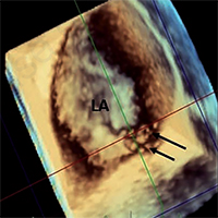
We report a case of a 55-year-old male admitted for cardiogenic embolic ischemic stroke work up. A transesophageal (TE) echocardiography (E) with contrast study to rule out patent foramen ovale (PFO) was performed; two-dimensional (2D) analysis did not detect any bubbles passage during Valsalva manoeuvre in the standard 2D cross sectional planes; further real time three-dimensional (3D) TEE imaging revealed passage of bubbles in the left atrium (LA) by both real-time 3DTEE imaging and by the 2D unconventional cross-sectional planes allowed by 3DTEE imaging. Even though 2DTEE is considered to be the gold standard modality for diagnosing PFO, it has some limitations. It has never been reported about usefulness of 3DTEE in PFO imaging. Even in the presence of only a report, our case suggests that 3DE could have an additional value and will compliment 2D imaging in PFO assessment.
Downloads
Citations


