
A case report on expanding horizon of endobronchial ultrasound through esophagus
All claims expressed in this article are solely those of the authors and do not necessarily represent those of their affiliated organizations, or those of the publisher, the editors and the reviewers. Any product that may be evaluated in this article or claim that may be made by its manufacturer is not guaranteed or endorsed by the publisher.
Accepted: 22 May 2020
Authors
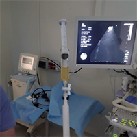
Endobronchial ultrasound has revolutionized the field of bronchoscopy and has become one of the most important tools for the diagnosis of intrathoracic lymphadenopathy and para-bronchial structures. The reach of this technique has not been limited to these structures and pleural lesions have been at times accessible. To our knowledge, pleural fluid collections have not been accessed with endobronchial ultrasound (EBUS) through oesophageal approach and rationale behind using this approach. We report a case of 70 years old man who has been referred from physician for the EBUS in view of hilar mass with mediastinal lymphadenopathy with pleural effusion. The endobronchial ultrasound through oesophagus (EUS-B) was done for thoracocentesis and lymph node cytology evaluation and ultimately endobronchial biopsy of hilar mass was done as rapid on-site (ROSE) analysis of lymph node was suggestive of necrotic tissue. The cytology report of lymph node and pleural effusion was positive for malignant cells. The final diagnosis was metastatic poorly differentiating adeno-squamous carcinoma.
Downloads
Citations


