Clinico-radiological and bronchoscopic predictors of microbiological yield in sputum negative tuberculosis in Pakistan

Submitted: June 14, 2021
Accepted: October 5, 2021
Published: December 6, 2021
Abstract Views: 1505
PDF: 396
Publisher's note
All claims expressed in this article are solely those of the authors and do not necessarily represent those of their affiliated organizations, or those of the publisher, the editors and the reviewers. Any product that may be evaluated in this article or claim that may be made by its manufacturer is not guaranteed or endorsed by the publisher.

Authors

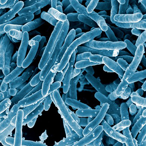
To determine association of clinico-radiological factors and radiological activity with diagnostic yield in sputum-smear negative tuberculosis (TB). Prospective observational study in the Military Hospital Rawalpindi (Pakistan) from July to December 2018. Adult patients having no contraindications to bronchoscopy were included. HIV positive patients and those on anti-tuberculosis therapy for more than one week were excluded. High-resolution computed tomography (HRCT) findings were classified based on active and inactive tuberculosis features. Washings were sent for acid-fast bacillus (AFB) smear, GeneXpert assay and cultures. Out of 215 patients, 42.3% (91) were diagnosed with microbiological or histological evidence of TB. On univariate analysis, cavitation (p-value <0.001), soft-tissue nodules (p-value 0.04), and endobronchial mucosal changes (p-value 0.02) were associated with culture positivity. Presence of cavitation (OR= 4.10; CI= 2.18,7.73; p-value<0.001) was the only independent predictor of microbiological yield. Diagnostic yield was 70%, 50%, 12.5% and 8.6% in patients with definitely active, probably active, indeterminate and inactive tuberculosis HRCT features respectively. Sensitivity, specificity, positive predictive value and negative predictive value of HRCT active TB were 95.38% (95% CI 87.10-99.04), 48.00 % (95% CI 39.78-56.30), 44.29% (95% CI 40.31-48.33), 96.00 % (95%CI 88.70-98.66) respectively. There was no significant association between age groups, smoking status and gender with diagnosis of tuberculosis in our study. Radiological activity and certain visualized bronchoscopic changes were associated with good diagnostic performance and can be used as predictive factors in diagnosis of active smear negative tuberculosis.  

Dimensions

Altmetric

PlumX Metrics

Downloads

Download data is not yet available.

Citations

World Health Organization. Global Tuberculosis Report 2018. Available from: https://apps.who.int/iris/handle/10665/274453
Raviglione M, Sulis G. Tuberculosis 2015: burden, challenges and strategy for control and elimination. Infect Dis Rep 2016;8:33–7. DOI: https://doi.org/10.4081/idr.2016.6570
Alsowey AM, Amin MI, Said AM. The predictive value of multidetector high resolution computed tomography in evaluation of suspected sputum smear negative active pulmonary tuberculosis in Egyptian Zagazig University Hospital Patients. Polish J Radiol 2017;82:808–16. DOI: https://doi.org/10.12659/PJR.903743
Tostmann A, Kik S V., Kalisvaart NA, et al. Tuberculosis transmission by patients with smear-negative pulmonary tuberculosis in a large cohort in the Netherlands. Clin Infect Dis 2008;47:1135–42. DOI: https://doi.org/10.1086/591974
Behr MA, Warren SA, Salamon H, et al. Transmission of Mycobacterium tuberculosis from patients smear-negative for acid-fast bacilli. Lancet 1999;353:444–9. DOI: https://doi.org/10.1016/S0140-6736(98)03406-0
Prasad R, Singh A. Role of bronchoscopy in diagnosis of smear-negative pulmonary tuberculosis. Egypt J Bronchol 2019;131-5.
Ji L, Lou YL, Wu ZX, et al. Usefulness of interferon-γ release assay for the diagnosis of sputum smear-negative pulmonary and extra-pulmonary TB in Zhejiang Province, China. Infect Dis Poverty 2017;6:1–5. DOI: https://doi.org/10.1186/s40249-017-0331-1
Zaidi SMA, Habib SS, Van Ginneken B, et al. Evaluation of the diagnostic accuracy of Computer-aided detection of tuberculosis on chest radiography among private sector patients in Pakistan. Sci Rep 2018;8:1–9. DOI: https://doi.org/10.1038/s41598-018-30810-1
Mondoni M, Repossi A, Carlucci P, et al. Bronchoscopic techniques in the management of patients with tuberculosis. Int J Infect Dis 2017;64:27–37. DOI: https://doi.org/10.1016/j.ijid.2017.08.008
Majmudar DK, Rajput DK. Role of HRCT in diagnosing disease activity of pulmonary tuberculosis. Int J Contemp Med Res 2017;4:1724–7.
Bhalla A, Goyal A, Guleria R, et al. Chest tuberculosis: Radiological review and imaging recommendations. Indian J Radiol Imaging 2015;25:213. DOI: https://doi.org/10.4103/0971-3026.161431
Ko Y, Lee HY, Park YB, et al. Correlation of microbiological yield with radiographic activity on chest computed tomography in cases of suspected pulmonary tuberculosis. PLoS One 2018;13:e0201748. DOI: https://doi.org/10.1371/journal.pone.0201748
Shaarrawy H, Zeidan M, Nasr A, et al. Assessment of the role of high resolution computed tomography in the diagnosis of suspected sputum smear negative active pulmonary TB. Egypt J Chest Dis Tuberc 2013;62:263–8. DOI: https://doi.org/10.1016/j.ejcdt.2013.05.006
Çalişkan T, Ozkisa T, Aribal S, et al. High resolution computed tomography findings in smear-negative pulmonary tuberculosis patients according to their culture status. J Thorac Dis 2014;6:706–12.
Shin JA, Chang YS, Kim TH, et al. Fiberoptic bronchoscopy for the rapid diagnosis of smear-negative pulmonary tuberculosis. BMC Infect Dis 2012;12:141. DOI: https://doi.org/10.1186/1471-2334-12-141
Le Palud P, Cattoir V, Malbruny B, et al. Retrospective observational study of diagnostic accuracy of the Xpert® MTB/RIF assay on fiberoptic bronchoscopy sampling for early diagnosis of smear-negative or sputum-scarce patients with suspected tuberculosis. BMC Pulm Med 2014;14:137. DOI: https://doi.org/10.1186/1471-2466-14-137
Lee JY, Lee KS, Jung KJ, et al. Pulmonary tuberculosis: CT and pathologic correlation. J Comput Assist Tomogr 2000;24:691–8. DOI: https://doi.org/10.1097/00004728-200009000-00005
To KW, Kam KM, Chan DPC, et al. Utility of GeneXpert in analysis of bronchoalveolar lavage samples from patients with suspected tuberculosis in an intermediate-burden setting. J Infect 2018;77:296–301. DOI: https://doi.org/10.1016/j.jinf.2018.06.011
Shimon G, Yonit W-W, Gabriel I, et al. The “tree-in-bud” pattern on chest CT: Radiologic and microbiologic correlation. Lung 2015;193:823–9. DOI: https://doi.org/10.1007/s00408-015-9759-x
Desai SR, Edey AJ, Hansell DM, et al. Morphologic predictors of a microbiological yield in patients with a tree-in-bud pattern on computed tomography. J Thorac Imaging 2014;29:240–5. DOI: https://doi.org/10.1097/RTI.0000000000000078
Nakanishi M, Demura Y, Ameshima S, et al. Utility of high-resolution computed tomography for predicting risk of sputum smear-negative pulmonary tuberculosis. Eur J Radiol 2010;73:545–50. DOI: https://doi.org/10.1016/j.ejrad.2008.12.009
Raghuvanshi V, Gopal Sood R, Jhobta A, et al. Use of high-resolution computed tomography (HRCT) in diagnosis of sputum negative pulmonary tuberculosis. Turkish Thorac J 2016;17:59–64. DOI: https://doi.org/10.5578/ttj.17.2.012
Haque S, Quaiser S, Agarwal A, et al. Fiberoptic bronchoscopy, as a valuable diagnostic option in sputum negative pulmonary tuberculosis: A prospective study. Int J Appl Basic Med Res 2012;2:123. DOI: https://doi.org/10.4103/2229-516X.106355
Shankar M, Saha KK, Sneha K, et al. Utility of fibreoptic bronchoscopy: An experience from a tertiary health care centre of Bihar , India. Int J Contemp Med Res 2018;5:1–4.
S C, Bo T, S K, et al. Outcome of fiber optic bronchoscopy in sputum smear negative pulmonary tuberculosis. Panacea J Med Sci 2015;5:33–9.
Sharma R, Sahasrabuddhe T, Showkat M, et al. A prospective study to evaluate the utility of bronchoalveolar lavage by fiberoptic bronchoscopy in sputum smear negative patients with high suspicion of pulmonary tuberculosis. Med J Dr DY Patil Univ 2012;5:43. DOI: https://doi.org/10.4103/0975-2870.97511
Ozkaya S, Bilgin S, Findik S, et al. Endobronchial tuberculosis: histopathological subsets and microbiological results. Multidiscip Respir Med 2012;7:34. DOI: https://doi.org/10.4081/mrm.2012.621
Şimşek A, Yapıcı İ, Babalık M, et al. Bronchoscopic diagnostic procedures and microbiological examinations in proving endobronchial tuberculosis. J Bras Pneumol 2016;42:191–5. DOI: https://doi.org/10.1590/S1806-37562015000000134
Peposhi IP, Kapisyzi PQ, Hafizi HS, et al. Value of bronchoscopy in the diagnosis of sputum smear negative pulmonary tuberculosis. Int J Res Med Sci 2017;5:3393 DOI: https://doi.org/10.18203/2320-6012.ijrms20173529
Lee J-J, Chong P-Y, Lin C-B, et al. High resolution chest CT in patients with pulmonary tuberculosis: Characteristic findings before and after antituberculous therapy. Eur J Radiol 2008;67:100–4. DOI: https://doi.org/10.1016/j.ejrad.2007.07.009
Pinto LM, Pai M, Dheda K, et al. Scoring systems using chest radiographic features for the diagnosis of pulmonary tuberculosis in adults: a systematic review. Eur Respir J 2013;42:480–94. DOI: https://doi.org/10.1183/09031936.00107412
Tozkoparan E, Deniz O, Ciftci F, et al. The roles of HRCT and clinical parameters in assessing activity of suspected smear negative pulmonary tuberculosis. Arch Med Res 2005;36:166–70. DOI: https://doi.org/10.1016/j.arcmed.2004.12.010

How to Cite

Ejaz, Taymmia, Mahmood Iqbal Malik, Jamal Ahmed, Rizwan Azam, Yousaf Jamal, and Sheema Saadia. 2021. “Clinico-Radiological and Bronchoscopic Predictors of Microbiological Yield in Sputum Negative Tuberculosis in Pakistan”. Monaldi Archives for Chest Disease 92 (3). https://doi.org/10.4081/monaldi.2021.1976.