Lung ultrasound: a narrative review and proposed protocol for patients admitted to Cardiac Rehabilitation Unit

Submitted: December 28, 2020
Accepted: June 26, 2021
Published: August 23, 2021
Abstract Views: 1459
PDF: 770
Publisher's note
All claims expressed in this article are solely those of the authors and do not necessarily represent those of their affiliated organizations, or those of the publisher, the editors and the reviewers. Any product that may be evaluated in this article or claim that may be made by its manufacturer is not guaranteed or endorsed by the publisher.

Authors

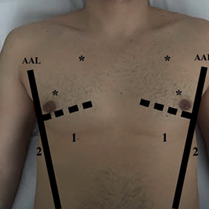
Lung ultrasonography (LUS) has become in the last 10 years a technique that has reduced the need of second level diagnostic methods such as chest X-ray (CXR) and computerize tomography (CT) for the diagnostic imaging of lung and pleural space, throughout its diagnostic accuracy, radiation free, low cost, real time and bedside approach. The common use of LUS has been recently extend to cardiac and pulmonary disease even in context of Cardiac Rehabilitation Unit and it could be an additional tool for physiotherapist for the management of patients during Rehabilitation course. The authors performed a literature review in PubMed and suggested a new standardize protocol for LUS, based on guidelines and expert consensus document, for patients admitted to Cardiac Rehabilitation Unit. In this protocol, LUS should be performed in six scan each hemithorax, covering twelve imagine regions. For each scan will be noted a specific physiologic or pathological patterns. Furthermore, we suggest for each patient, the use of the Lung Ultrasound Score (LUS score) to obtain a global view of lung aeration and to monitor any changes during the hospitalization. An increase in score range indicates a more severe condition. This Lung Ultrasonography Protocol should be performed in all patients at the time of admission to Cardiac Rehabilitation Unit to monitoring the aeration of the lungs and the possible lung and/or pleura complications after a cardiac disease avoiding the use of second level surveys.

Dimensions

Altmetric

PlumX Metrics

Downloads

Download data is not yet available.

Citations

Oks M, Cleven KL, Cardenas‐Garcia J, et al. The effect of point‐of‐care ultrasonography on imaging studies in the medical ICU: a comparative study. Chest 2014;146:1574-7. DOI: https://doi.org/10.1378/chest.14-0728
Mayo PH, Copetti R, Feller-Kopman D, et al. Thoracic ultrasonography: a narrative review. Intensive Care Med 2019; 45:1200-11. DOI: https://doi.org/10.1007/s00134-019-05725-8
Mancusi C, Carlino MV, Sforza A. Point-of-care ultrasound with pocket-size devices in emergency department. Echocardiography 2019;36:1755-64. DOI: https://doi.org/10.1111/echo.14451
Sforza A, Mancusi C, Carlino MV, et al. Diagnostic performance of multi-organ ultrasound with pocket-sized device in the management of acute dyspnea. Cardiovasc Ultrasound 2017;15:16. DOI: https://doi.org/10.1186/s12947-017-0105-8
Carlino MV, Paladino F, Sforza A, et al. Assessment of left atrial size in addition to focused cardiopulmonary ultrasound improves diagnostic accuracy of acute heart failure in the Emergency Department. Echocardiography 2018;35:785-79. DOI: https://doi.org/10.1111/echo.13851
Gargani L, Volpicelli G. How I do it: lung ultrasound. Cardiovasc Ultrasound 2014;12:25. DOI: https://doi.org/10.1186/1476-7120-12-25
Sforza A, Carlino MV, Guarino M, et al. Anterior vs lateral symmetric interstitial syndrome in the diagnosis of acute heart failure. Int J Cardiol 2019; 280:130-2. DOI: https://doi.org/10.1016/j.ijcard.2019.01.013
Sforza A, Carlino MV, Guarino M, et al. A case of pulmonary edema: The critical role of lung-heart integrated ultrasound examination. Monaldi Arch Chest Dis 2018;88:982. DOI: https://doi.org/10.4081/monaldi.2018.982
Lichtenstein DA, Mezière GA. Relevance of lung ultrasound in the diagnosis of acute respiratory failure: the BLUE protocol. Chest 2008;134:117-25. DOI: https://doi.org/10.1378/chest.07-2800
Lichtenstein DA. BLUE-protocol and FALLS-protocol: two applications of lung ultrasound in the critically ill. Chest 2015;147:1659-70. DOI: https://doi.org/10.1378/chest.14-1313
Lichtenstein D. Novel approaches to ultrasonography of the lung and pleural space: where are we now? Breathe Sheff Engl 2017;13:100-11. DOI: https://doi.org/10.1183/20734735.004717
Soldati G, Smargiassi A, Mariani AA, et al. Novel aspects in diagnostic approach to respiratory patients: is it the time for a new semiotics? Multidiscip Respir Med 2017;12:15. DOI: https://doi.org/10.4081/mrm.2017.242
Soldati G, Demi M, Smargiassi A, et al. The role of ultrasound lung artifacts in the diagnosis of respiratory diseases. Expert Rev Respir Med 2019;13:163-72. DOI: https://doi.org/10.1080/17476348.2019.1565997
Mohanty K, Blackwell J, Egan T, et al. Characterization of the lung parenchyma using ultrasound multiple scattering. Ultrasound Med Biol 2017; 43:993-1003. DOI: https://doi.org/10.1016/j.ultrasmedbio.2017.01.011
Dietrich CF, Mathis G, Blaivas M, et al. Lung B‐line artefacts and their use. J Thorac Dis 2016;8:1356-65. DOI: https://doi.org/10.21037/jtd.2016.04.55
Francisco MJ Neto, Rahal A Junior, Vieira FA, et al. Advances in lung ultrasound. Einstein (Sao Paulo) 2016;14:443-8. DOI: https://doi.org/10.1590/S1679-45082016MD3557
Francis Chun Yue Lee. The curtain sign in lung ultrasound. J Med Ultrasound 2017;25:101-4. DOI: https://doi.org/10.1016/j.jmu.2017.04.005
Pedretti RFE, Fattirolli F, Griffo R, et al. Cardiac prevention and rehabilitation "3.0": from acute to chronic phase. Position paper of the Italian association for cardiovascular prevention and rehabilitation (GICR-IACPR). Monaldi Arch Chest Dis 2018;88:1004. DOI: https://doi.org/10.4081/monaldi.2018.1004
Prabhu NV, Maiya AG, Prabhu NS. Impact of cardiac rehabilitation on functional capacity and physical activity after coronary revascularization: a scientific review. Cardiol Res Pract 2020; 2020:1236968. DOI: https://doi.org/10.1155/2020/1236968
Brogi E, Bignami E, Sidoti A, et al. Could the use of bedside lung ultrasound reduce the number of chest x‐rays in the intensive care unit? Cardiovasc Ultrasound 2017;15:23. DOI: https://doi.org/10.1186/s12947-017-0113-8
Platz E, Jhund PS, Girerd N, et al. Study group on acute heart failure of the acute cardiovascular care association and the heart failure association of the European society of cardiology. Expert consensus document: reporting checklist for quantification of pulmonary congestion by lung ultrasound in heart failure. Eur J Heart Fail.2019;21:844-85. DOI: https://doi.org/10.1002/ejhf.1499
Martindale JL, Wakai A, Collins SP, et al. Diagnosing acute heart failure in the emergency department: a systematic review and meta-analysis. Acad Emerg Med 2016;23:223-42. DOI: https://doi.org/10.1111/acem.12878
Soldati G, Demi M. The use of lung ultrasound images for the differential diagnosis of pulmonary and cardiac interstitial pathology. J Ultrasound 2017; 20:91-6. DOI: https://doi.org/10.1007/s40477-017-0244-7
Pivetta E, Goffi A, Nazerian P, et al. Study group on lung ultrasound from the Molinette and Careggi Hospitals. Lung ultrasound integrated with clinical assessment for the diagnosis of acute decompensated heart failure in the emergency department: a randomized controlled trial. Eur J Heart Fail 2019; 21:754-66. DOI: https://doi.org/10.1002/ejhf.1379
Pivetta E, Goffi A, Lupia E, et al. SIMEU group for lung ultrasound in the emergency department in Piedmont. Lung ultrasound-implemented diagnosis of acute decompensated heart failure in the ED: a SIMEU multicenter study. Chest 2015;148:202-10 DOI: https://doi.org/10.1378/chest.14-2608
Ponikowski P, Voors AA, Anker SD, et al. ESC guidelines for the diagnosis and treatment of acute and chronic heart failure: the Task Force for the Diagnosis and Treatment of Acute and Chronic Heart Failure of the European Society of Cardiology. Eur J Heart Fail 2016;18:891-975. DOI: https://doi.org/10.1002/ejhf.592
Efremov SM, Kuzkov VV, Fot EV, et al. Lung ultrasonography and cardiac surgery: A narrative review. J Cardiothorac Vasc Anesth 2020;34:3113-24. DOI: https://doi.org/10.1053/j.jvca.2020.01.032
Brogi E, Gargani L, Bignami E, et al. Thoracic ultrasound for pleural effusion in the intensive care unit: a narrative review from diagnosis to treatment. Crit Care 2017;21:325. DOI: https://doi.org/10.1186/s13054-017-1897-5
Balik M, Plasil P, Waldauf P, et al. Ultrasound estimation of volume of pleural fluid in mechanically ventilated patients. Intensive Care Med 2006;32:318. DOI: https://doi.org/10.1007/s00134-005-0024-2
Picano E, Scali MC, Ciampi Q, Lichtenstein D. Lung ultrasound for the cardiologist. JACC Cardiovasc Imaging 2018;11:1692-705. DOI: https://doi.org/10.1016/j.jcmg.2018.06.023
Lazarevic A, Dobric M, Goronja B, et al. Lung ultrasound-guided therapeutic thoracentesis in refractory congestive heart failure. Acta Cardiol 2020;75:398-405. DOI: https://doi.org/10.1080/00015385.2019.1591677
Domokos D, SzaboA, Banhegyi G, et al. Needle aspiration for treating iatrogenic pneumothorax after cardiac electronic device implantation: a pilot study. J Interv Card Electrophysiol 2020;57:295-301. DOI: https://doi.org/10.1007/s10840-019-00596-x
Alrajab S, Youssef AM, Akkus NI, Caldito G. Pleural ultrasonography versus chest radiography for the diagnosis of pneumothorax: review of the literature and meta‐analysis. Crit Care 2013;17:R208. DOI: https://doi.org/10.1186/cc13016
Volpicelli G. Sonographic diagnosis of pneumothorax. Intensive Care Med 2011;37:224-32. DOI: https://doi.org/10.1007/s00134-010-2079-y
Santos-Silva J, Lichtenstein D, Tuinman PR, Elbers PWG. The lung point, still a sign specific to pneumothorax. Intensive Care Med 2019;45:1327-8. DOI: https://doi.org/10.1007/s00134-019-05677-z
Lichtenstein DA, Menu Y. A bedside ultrasound sign ruling out pneumothorax in the critically ill: lung sliding. Chest 1995;108:1345-8. DOI: https://doi.org/10.1378/chest.108.5.1345
Alzahrani SA, Al-Salamah MA, Al-Madani WH, et al. Systematic review and meta-analysis for the use of ultrasound versus radiology in diagnosing of pneumonia. Crit Ultrasound J 2017;9:6. DOI: https://doi.org/10.1186/s13089-017-0059-y
Reissig A, Copetti R, Mathis G et al. Lung ultrasound in the diagnosis and follow-up of community-acquired pneumonia: a prospective, multicenter, diagnostic accuracy study. Chest 2012;142:965-72. DOI: https://doi.org/10.1378/chest.12-0364
Soldati G, Copetti R. Thoracic ultrasound. Turin, Edizioni medico-scientifiche; 2017.
Montero Baladía M, Arroyo Diez M, Badallo Areválo O. Basic sign in lung echography. Liquid and air bronchogram: differentiation. Med Intensiva 2018;42:19. DOI: https://doi.org/10.1016/j.medine.2018.07.004
Asano M, Watanabe H, Sato K et al. Validity of ultrasound lung comets for assessment of the severity of interstitial pneumonia. J Ultrasound Med 2018; 37:1523-31. DOI: https://doi.org/10.1002/jum.14497
Volpicelli G, Elbarbary M, Blaivas M, et al. International evidence‐based recommendations for point‐of‐care lung ultrasound. Intensive Care Med 2012;38:577–91. DOI: https://doi.org/10.1007/s00134-012-2513-4
McKiernan S, Chiarelli P, Warren-Forward H. Diagnostic ultrasound use in physiotherapy, emergency medicine, and anaesthesiology. Radiography 2010;16:154-9. DOI: https://doi.org/10.1016/j.radi.2009.12.004
Mojoli F, Bouhemad B, Mongodi S, Lichtenstein D. Lung ultrasound for critically ill patients. Am J Respir Crit Care Med 2019;199:701-14. DOI: https://doi.org/10.1164/rccm.201802-0236CI
Crimi C, Heffler E, Augelletti T, et al. Utility of ultrasound assessment of diaphragmatic function before and after pulmonary rehabilitation in COPD patients. Int J Chron Obstruct Pulmon Dis 2018;13:3131-9. DOI: https://doi.org/10.2147/COPD.S171134
Karthika M, Wong D, Nair SG, et al. Lung ultrasound: the emerging role of respiratory therapists. Respir Care 2019;64:217-29. DOI: https://doi.org/10.4187/respcare.06179
Le Neindre A, Mongodi S, Philippart F, Bouhemad B. Thoracic ultrasound: potential new tool for physiotherapists in respiratory management. A narrative review. J Crit Care 2016;31:101-9. DOI: https://doi.org/10.1016/j.jcrc.2015.10.014
Hayward S, Duncan C. Physiotherapists use of lung ultrasound during the COVID-19 pandemic: a practical guideline on supporting acute hospital colleagues. Association of Chartered Physiotherapists in Respiratory Care; 2020.
Hayward S, Kelly D. Respiratory physiotherapy and lung ultrasound: a service evaluation of a training programme. J Assoc Chart Physiother Respir Care 2017; 50:51-58.
Soummer A, Perbet S, Brisson H, et al. Ultrasound assessment of lung aeration loss during a successful weaning trial predicts postextubation distress. Crit Care Med 2012;40:2064-72. DOI: https://doi.org/10.1097/CCM.0b013e31824e68ae

How to Cite

Bertolone, Dario Tino, Cristina De Colle, Francesco Rozza, Ilaria Fucile, Ciro Santoro, Maurizio Conte, Nicola De Luca, and Costantino Mancusi. 2021. “Lung Ultrasound: A Narrative Review and Proposed Protocol for Patients Admitted to Cardiac Rehabilitation Unit”. Monaldi Archives for Chest Disease 92 (1). https://doi.org/10.4081/monaldi.2021.1753.