
Pneumology - Reviews
Vol. 91 No. 2 (2021)
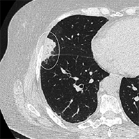
Imaging the COVID-19: a practical guide
Publisher's note
All claims expressed in this article are solely those of the authors and do not necessarily represent those of their affiliated organizations, or those of the publisher, the editors and the reviewers. Any product that may be evaluated in this article or claim that may be made by its manufacturer is not guaranteed or endorsed by the publisher.
All claims expressed in this article are solely those of the authors and do not necessarily represent those of their affiliated organizations, or those of the publisher, the editors and the reviewers. Any product that may be evaluated in this article or claim that may be made by its manufacturer is not guaranteed or endorsed by the publisher.
Received: 2 October 2020
Accepted: 17 January 2021
Accepted: 17 January 2021
2650
Views
1405
Downloads
Authors
The Coronavirus Disease 2019 (COVID-19) represents the first medical catastrophe of the new millennium. Although imaging is not a screening test for COVID-19, it plays a crucial role in evaluation and follow-up of COVID-19 patients. In this paper, we will review typical and atypical imaging findings of COVID-19.
Downloads
Download data is not yet available.
Citations


Chan JF-W, Yuan S, Kok K-H, et al. A familial cluster of pneumonia associated with the 2019 novel coronavirus indicating person-to-person transmission: a study of a family cluster. Lancet (London, England). 2020;395(10223):514-523. DOI: https://doi.org/10.1016/S0140-6736(20)30154-9
Huang C, Wang Y, Li X, et al. Clinical features of patients infected with 2019 novel coronavirus in Wuhan, China. Lancet (London, England). 2020;395(10223):497-506. DOI: https://doi.org/10.1016/S0140-6736(20)30183-5
Zhu N, Zhang D, Wang W, et al. A Novel Coronavirus from Patients with Pneumonia in China, 2019. N Engl J Med. 2020;382(8):727-733. DOI: https://doi.org/10.1056/NEJMoa2001017
Wang F-S, Zhang C. What to do next to control the 2019-nCoV epidemic? Lancet (London, England). 2020;395(10222):391-393. DOI: https://doi.org/10.1016/S0140-6736(20)30300-7
World Health Organization. World Health Organization (2020) - Coronavirus disease (COVID-19) Situation Report-135. https://www.who.int/docs/default-source/coronaviruse/situation-reports/20200604-covid-19-sitrep-136.pdf?sfvrsn=fd36550b_2.
Yang X, Yu Y, Xu J, et al. Clinical course and outcomes of critically ill patients with SARS-CoV-2 pneumonia in Wuhan, China: a single-centered, retrospective, observational study. Lancet Respir Med. 2020;8(5):475-481. DOI: https://doi.org/10.1016/S2213-2600(20)30079-5
Kimball A, Hatfield KM, Arons M, et al. Asymptomatic and Presymptomatic SARS-CoV-2 Infections in Residents of a Long-Term Care Skilled Nursing Facility - King County, Washington, March 2020. MMWR Morb Mortal Wkly Rep. 2020;69(13):377-381. DOI: https://doi.org/10.15585/mmwr.mm6913e1
Wang XF, Shi GC, Wan HY, et al. Clinical features of three avian influenza H7N9 virus-infected patients in Shanghai. Clin Respir J. 2014;8(4):410-416. DOI: https://doi.org/10.1111/crj.12087
Badawi A, Ryoo SG. Prevalence of comorbidities in the Middle East respiratory syndrome coronavirus (MERS-CoV): a systematic review and meta-analysis. Int J Infect Dis IJID Off Publ Int Soc Infect Dis. 2016;49:129-133. DOI: https://doi.org/10.1016/j.ijid.2016.06.015
Raptis CA, Hammer MM, Short RG, et al. Chest CT and Coronavirus Disease (COVID-19): A Critical Review of the Literature to Date. AJR Am J Roentgenol. April 2020:1-4. DOI: https://doi.org/10.2214/AJR.20.23202
Guan W-J, Ni Z-Y, Hu Y, et al. Clinical Characteristics of Coronavirus Disease 2019 in China. N Engl J Med. 2020;382(18):1708-1720. DOI: https://doi.org/10.1056/NEJMoa2002032
Qian M, Yi Q, Qihua F, Ming G. Understanding the influencing factors of nucleic acid detection of 2019 novel coronavirus. Chin J Lab Med. 2020;10.
World Health Organization. Infection prevention and control during health care when novel coronavirus (nCoV) infection is suspected. https://www.who.int/publications/i/item/10665-331495.
Rubin GD, Ryerson CJ, Haramati LB, et al. The Role of Chest Imaging in Patient Management during the COVID-19 Pandemic: A Multinational Consensus Statement from the Fleischner Society. Radiology. 2020;296(1):172-180. DOI: https://doi.org/10.1148/radiol.2020201365
Li Y, Xia L. Coronavirus Disease 2019 (COVID-19): Role of Chest CT in Diagnosis and Management. AJR Am J Roentgenol. 2020;214(6):1280-1286.
Mossa-Basha M, Medverd J, Linnau K, et al. Policies and Guidelines for COVID-19 Preparedness: Experiences from the University of Washington. Radiology. April 2020:201326. DOI: https://doi.org/10.1148/radiol.2020201326
American College of Radiology (ACR). ACR Recommendations for the use of Chest Radiography and Computed Tomography (CT) for Suspected COVID-19 Infection. https://www.acr.org/Advocacy-and-Economics/ACR-Position-Statements/Recommendations-for-Chest-Radiography-and-CT-for-Suspected-COVID19-Infection.
Chen S-G, Chen J-Y, Yang Y-P, Chien C-S, Wang M-L, Lin L-T. Use of Radiographic Features in COVID-19 Diagnosis. J Chinese Med Assoc. 2020:1. DOI: https://doi.org/10.1097/JCMA.0000000000000336
Soldati G, Smargiassi A, Inchingolo R, et al. Is there a role for lung ultrasound during the COVID-19 pandemic? J Ultrasound Med. 2020:1-4. DOI: https://doi.org/10.1002/jum.15284
Smith MJ, Hayward SA, Innes SM, Miller ASC. Point-of-care lung ultrasound in patients with COVID-19 - a narrative review. Anaesthesia. April 2020. doi:10.1111/anae.15082 DOI: https://doi.org/10.1111/anae.15082
Xu B, Xing Y, Peng J, et al. Chest CT for Detecting COVID-19: A Systematic Review and Meta-Analysis of Diagnostic Accuracy. 2020;(866):14. DOI: https://doi.org/10.21203/rs.3.rs-20481/v1
Song F, Shi N, Shan F, et al. Emerging 2019 novel coronavirus (2019-NCoV) pneumonia. Radiology. 2020;295(1):210-217. DOI: https://doi.org/10.1148/radiol.2020200274
Pan F, Ye T, Sun P, et al. Time Course of Lung Changes On Chest CT During Recovery From 2019 Novel Coronavirus (COVID-19) Pneumonia. Radiology. 2020:200370. DOI: https://doi.org/10.1148/radiol.2020200370
Han R, Huang L, Jiang H, Dong J, Peng H, Zhang D. Early Clinical and CT Manifestations of Coronavirus Disease 2019 (COVID-19) Pneumonia. AJR Am J Roentgenol. 2020;(1):1-6. DOI: https://doi.org/10.2214/AJR.20.22961
Shi H, Han X, Zheng C. Evolution of CT Manifestations in a Patient Recovered from 2019 Novel Coronavirus (2019-nCoV) Pneumonia in Wuhan, China. Radiology. 2020;295(1):20. DOI: https://doi.org/10.1148/radiol.2020200269
Chung M, Bernheim A, Mei X, et al. CT Imaging Features of 2019 Novel Coronavirus (2019-nCoV). Radiology. 2020;295(1):202-207. DOI: https://doi.org/10.1148/radiol.2020200230
Ai T, Yang Z, Hou H, et al. Correlation of Chest CT and RT-PCR Testing in Coronavirus Disease 2019 (COVID-19) in China: A Report of 1014 Cases. Radiology. February 2020:200642. DOI: https://doi.org/10.1148/radiol.2020200642
Bai Y, Yao L, Wei T, et al. Presumed Asymptomatic Carrier Transmission of COVID-19. JAMA. 2020;323(14):1406-1407. DOI: https://doi.org/10.1001/jama.2020.2565
Meng H, Xiong R, He R, et al. CT imaging and clinical course of asymptomatic cases with COVID-19 pneumonia at admission in Wuhan, China. J Infect. 2020;(January). DOI: https://doi.org/10.1016/j.jinf.2020.04.004
Hansell DM, Bankier AA, MacMahon H, McLoud TC, Müller NL, Remy J. Fleischner Society: Glossary of terms for thoracic imaging. Radiology. 2008;246(3):697-722. DOI: https://doi.org/10.1148/radiol.2462070712
Salehi S, Abedi A, Balakrishnan S, Gholamrezanezhad A. Coronavirus Disease 2019 (COVID-19): A Systematic Review of Imaging Findings in 919 Patients. AJR Am J Roentgenol. 2020;(July):1-7. DOI: https://doi.org/10.2214/AJR.20.23034
Xu Z, Shi L, Wang Y, et al. Pathological findings of COVID-19 associated with acute respiratory distress syndrome. Lancet Respir Med. 2020;8(4):420-422. DOI: https://doi.org/10.1016/S2213-2600(20)30076-X
Hu Q, Guan H, Sun Z, et al. Early CT features and temporal lung changes in COVID-19 pneumonia in Wuhan, China. Eur J Radiol. 2020;128:109017. DOI: https://doi.org/10.1016/j.ejrad.2020.109017
Bernheim A, Mei X, Huang M, et al. Chest CT Findings in Coronavirus Disease-19 (COVID-19): Relationship to Duration of Infection. Radiology. February 2020:200463. DOI: https://doi.org/10.1148/radiol.2020200463
Caruso D, Zerunian M, Polici M, et al. Chest CT Features of COVID-19 in Rome, Italy. Radiology. April 2020:201237. DOI: https://doi.org/10.1148/radiol.2020201237
Parry AH, Wani AH. Segmental Pulmonary Vascular Changes in COVID-19 Pneumonia. AJR Am J Roentgenol. May 2020:W1. DOI: https://doi.org/10.2214/AJR.20.23443
Chiarenza A, Esposto Ultimo L, Falsaperla D, et al. Chest imaging using signs, symbols, and naturalistic images: a practical guide for radiologists and non-radiologists. Insights Imaging. 2019;10(1). DOI: https://doi.org/10.1186/s13244-019-0789-4
Farias L de PG de, Strabelli DG, Sawamura MVY. COVID-19 pneumonia and the reversed halo sign. J Bras Pneumol. 2020;46(2):e20200131. DOI: https://doi.org/10.36416/1806-3756/e20200131
Li Y, Xia L. Coronavirus Disease 2019 (COVID-19): Role of Chest CT in Diagnosis and Management. AJR Am J Roentgenol. 2020;(October):1-7. DOI: https://doi.org/10.2214/AJR.20.22954
Xia W, Shao J, Guo Y, Peng X, Li Z, Hu D. Clinical and CT features in pediatric patients with COVID-19 infection: Different points from adults. Pediatr Pulmonol. 2020;55(5):1169-1174. DOI: https://doi.org/10.1002/ppul.24718
Thannickal VJ, Toews GB, White ES, Lynch III JP, Martinez FJ. Mechanisms of Pulmonary Fibrosis. Annu Rev Med. 2004;55(1):395-417. DOI: https://doi.org/10.1146/annurev.med.55.091902.103810
Zhou S, Wang Y, Zhu T, Xia L. CT Features of Coronavirus Disease 2019 (COVID-19) Pneumonia in 62 Patients in Wuhan, China. AJR Am J Roentgenol. 2020;(October):1-8. DOI: https://doi.org/10.2214/AJR.20.22975
Li K, Wu J, Wu F, et al. The Clinical and Chest CT Features Associated with Severe and Critical COVID-19 Pneumonia. Invest Radiol. 2020;55(6):1-5. DOI: https://doi.org/10.1097/RLI.0000000000000672
Yu M, Xu D, Lan L, et al. Thin-section Chest CT Imaging of Coronavirus Disease 2019 Pneumonia: Comparison Between Patients with Mild and Severe Disease. Radiol Cardiothorac Imaging. 2020;2(2):e200126. DOI: https://doi.org/10.1148/ryct.2020200126
Zhu J, Zhong Z, Li H, et al. CT imaging features of 4,121 patients with COVID‐19: a meta‐analysis. J Med Virol. 2020;(April):1-12.
Wu J, Pan J, Teng D, Xu X, et al.. Interpretation of CT signs of 2019 novel coronavirus (COVID-19) pneumonia. Eur Radiol. 2020;(December 2019). doi:10.1007/s00330-020-06915-5 DOI: https://doi.org/10.1007/s00330-020-06915-5
Kanne JP, Little BP, Chung JH, Elicker BM, Ketai LH. Essentials for Radiologists on COVID-19: An Update-Radiology Scientific Expert Panel. Radiology. February 2020:200527.
Mao R, Qiu Y, He J, et al. Articles Manifestations and prognosis of gastrointestinal and liver involvement in patients with COVID-19 : a systematic review and meta-analysis. Lancet Gastroenterol Hepatol. 2020;1253(20). DOI: https://doi.org/10.1016/S2468-1253(20)30240-5
Siegel A, Chang PJ, Jarou ZJ, et al. Lung Base Findings of Coronavirus Disease (COVID-19) on Abdominal CT in Patients With Predominant Gastrointestinal Symptoms. Am J Roentgenol. 2020;(September):1-3. DOI: https://doi.org/10.2214/AJR.20.23232
Bhayana R, Som A, Li MD, et al. Abdominal Imaging Findings in COVID-19: Preliminary Observations. Radiology. May 2020:201908. DOI: https://doi.org/10.1148/radiol.2020201908
Chai X, Hu L, Zhang Y, et al. Specific ACE2 Expression in Cholangiocytes May Cause Liver Damage After 2019-nCoV Infection. bioRxiv. 2020:2020.02.03.931766. DOI: https://doi.org/10.1101/2020.02.03.931766
Xiao F, Tang M, Zheng X, Liu Y, Li X, Shan H. Evidence for Gastrointestinal Infection of SARS-CoV-2. Gastroenterology. 2020;158(6):1831-1833.e3. DOI: https://doi.org/10.1053/j.gastro.2020.02.055
de Barry O, Mekki A, Diffre C, Seror M, Hajjam M El, Carlier R-Y. Arterial and venous abdominal thrombosis in a 79-year-old woman with COVID-19 pneumonia. Radiol case reports. April 2020. DOI: https://doi.org/10.1016/j.radcr.2020.04.055
Beccara L, Pacioni C, Ponton S, Francavilla S, Cuzzoli A. of Case Reports in Arterial Mesenteric Thrombosis as a Complication of SARS-CoV-2 Infection of Case Reports in. 2020:4-6.
Mao L, Jin H, Wang M, et al. Neurologic Manifestations of Hospitalized Patients with Coronavirus Disease 2019 in Wuhan, China. JAMA Neurol. 2020. DOI: https://doi.org/10.1001/jamaneurol.2020.1127
Kandemirli SG, Dogan L, Sarikaya ZT, et al. Brain MRI Findings in Patients in the Intensive Care Unit with COVID-19 Infection. Radiology. May 2020:201697.
Asadi-Pooya AA, Simani L. Central nervous system manifestations of COVID-19: A systematic review. J Neurol Sci. 2020;413:116832. DOI: https://doi.org/10.1016/j.jns.2020.116832
Zanin L, Saraceno G, Panciani PP, et al. SARS-CoV-2 can induce brain and spine demyelinating lesions. Acta Neurochir (Wien). 2020;162(7):1491-1494. DOI: https://doi.org/10.1007/s00701-020-04374-x
Sachs JR, Gibbs KW, Swor DE, et al. COVID-19-Associated Leukoencephalopathy. Radiology. May 2020:201753. DOI: https://doi.org/10.1148/radiol.2020201753
How to Cite
“Imaging the COVID-19: A Practical Guide”. 2021. Monaldi Archives for Chest Disease 91 (2). https://doi.org/10.4081/monaldi.2021.1630.
Copyright (c) 2021 The Author(s)


This work is licensed under a Creative Commons Attribution-NonCommercial 4.0 International License.