
Pneumology - Case Reports
28 September 2021
Vol. 92 No. 1 (2022)
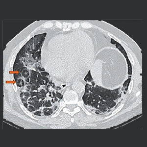
Bull’s eye sign – A diagnostic clinch in COVID-19 pneumonia
Publisher's note
All claims expressed in this article are solely those of the authors and do not necessarily represent those of their affiliated organizations, or those of the publisher, the editors and the reviewers. Any product that may be evaluated in this article or claim that may be made by its manufacturer is not guaranteed or endorsed by the publisher.
All claims expressed in this article are solely those of the authors and do not necessarily represent those of their affiliated organizations, or those of the publisher, the editors and the reviewers. Any product that may be evaluated in this article or claim that may be made by its manufacturer is not guaranteed or endorsed by the publisher.
1462
Views
426
Downloads
Authors
Although typical imaging findings of COVID-19 pneumonia has been described it may be difficult at times to distinguish it from other viral pneumonias. In the following case series, we describe a typical sign i.e. Bull’s-eye sign in COVID-19 pneumonia. As this sign is not associated with any known pulmonary disease, so its presence may help radiologists to differentiate COVID-19 pneumonia from its mimics.
Altmetrics
Downloads
Download data is not yet available.
Citations


Huang C, Wang Y, Li X, et al. Clinical features of patients infected with 2019 novel coronavirus in Wuhan, China. Lancet 2020;395:497-506. DOI: https://doi.org/10.1016/S0140-6736(20)30183-5
Song F, Shi N, Shan F, et al. Emerging coronavirus 2019-nCoV pneumonia. Radiology 2020;295:210-7. DOI: https://doi.org/10.1148/radiol.2020200274
Pan F, Ye T, Sun P, et al. Time course of lung changes on chest CT during recovery from 2019 novel Coronavirus (COVID-19) pneumonia. Radiology 2020;295:715–21. DOI: https://doi.org/10.1148/radiol.2020200370
Chung M, Bernheim A, Mei X, et al. CT imaging features of 2019 novel coronavirus (2019-nCoV). Radiology 2020;295:202–7. DOI: https://doi.org/10.1148/radiol.2020200230
Ai T, Yang Z, Hou H, et al. Correlation of chest CT and RT-PCR testing in coronavirus disease 2019 (COVID-19) in China: a report of 1014 cases. Radiology 2020;296:E32–E40. DOI: https://doi.org/10.1148/radiol.2020200642
Kong W, Agarwal PP. Chest imaging appearance of COVID-19 infection. Radiol Cardiothorac Imaging 2020;2:e200028. DOI: https://doi.org/10.1148/ryct.2020200028
Yoon SH, Lee KH, Kim JY, et al. Chest radiographic and CT findings of the 2019 novel coronavirus disease (COVID-19): analysis of nine patients treated in Korea. Korean J Radiol 2020;21:494–500. DOI: https://doi.org/10.3348/kjr.2020.0132
Baque-Juston M, Pellegrin A, Leroy S, et al. Organizing pneumonia: what is it? A conceptual approach and pictorial review. Diagn Interv Imaging 2014;95:771-7. DOI: https://doi.org/10.1016/j.diii.2014.01.004
Bernheim A, Mei X, Huang M, et al. Chest CT findings in coronavirus disease-19 (COVID-19): relationship to duration of infection. Radiology 2020;295:685–91. DOI: https://doi.org/10.1148/radiol.2020200463
Li Y, Xia L. Coronavirus disease 2019 (COVID-19): role of chest CT in diagnosis and management. AJR Am J Roentgenol 2020;214:1280-6. DOI: https://doi.org/10.2214/AJR.20.22954
Lomoro P,Verde F, Zerboni F, et al. COVID-19 pneumonia manifestations at the admission on chest ultrasound, radiographs, and CT: single-center study and comprehensive radiologic literature review. Eur J Radiol Open 2020;7:1–11. DOI: https://doi.org/10.1016/j.ejro.2020.100231
Bai HX, Hsieh B, Xiong Z, et al. Performance of radiologists in differentiating COVID-19 from viral pneumonia on chest CT. Radiology 2020;296:E46-E54. DOI: https://doi.org/10.1148/radiol.2020200823
Zhao H, Liang T, Wu CC, Jin C. The reversed halo sign in COVID-19 pneumonia. Research Square 2020. Available from: https://www.researchsquare.com/article/rs-20394/v1
McLaren TA, Gruden JF, Green DB. The bullseye sign: A variant of the reverse halo sign in COVID-19 pneumonia. Clin Imaging 2020; 68:191-6. DOI: https://doi.org/10.1016/j.clinimag.2020.07.024
Bikdeli B, Madhavan MV, Jimenez D, et al. COVID-19 and thrombotic or thromboembolic disease: implications for prevention, antithrombotic therapy, and follow-up: JACC state-of-the-art review. J Am Coll Cardiol 2020;75:2950–73. DOI: https://doi.org/10.1016/j.jacc.2020.04.031
Boraschi P. COVID-19 pulmonary involvement: is really an interstitial pneumonia? Acad Radiol 2020;27:900. DOI: https://doi.org/10.1016/j.acra.2020.04.010
Wu Y, Xie Y, Wang X. Longitudinal CT findings in COVID-19 pneumonia: case presenting organizing pneumonia pattern. Radiol Cardiothorac Imaging 2020;2:e200031. DOI: https://doi.org/10.1148/ryct.2020200031
Shaghaghi S, Daskareh M, Irannejad M, et al. Target-shaped combined halo and reversed-halo sign, an atypical chest CT finding in COVID-19. Clin Imaging 2021;69:72-4. DOI: https://doi.org/10.1016/j.clinimag.2020.06.038
Supporting Agencies
NoneHow to Cite
“Bull’s Eye Sign – A Diagnostic Clinch in COVID-19 Pneumonia”. 2021. Monaldi Archives for Chest Disease 92 (1). https://doi.org/10.4081/monaldi.2021.1908.
Copyright (c) 2021 The Author(s)


This work is licensed under a Creative Commons Attribution-NonCommercial 4.0 International License.